Logo
Login Signup
images
MRI
DWI MRI: Bilateral white matter hyperintensities.
T2 MRI: Supratentorial white matter hyperintensities.
T2-FLAIR MRI: Periventricular white matter hyperintensities sparing U-fibers.
Histopathology
H&E ×200: White matter vacuoles & astrogliosis.
H&E ×400: Vacuolization & microglial activation.
View Discussion
  • Title: Toxic Leukoencephalopathy in a 45-year-old Female Opioid User
  • Diagnosis Title: Toxic Leukoencephalopathy due to Opioid Use (Heroin/Morphine/Oxycodone)
  • Disease Specialty: Neurology
  • Ease of Diagnosis: Difficult
  • Certainty: Certain
  • Ethnicity: Asian
  • Segment: Adult Female
  • Image Quality: Medium
  • Clinical Examination: Patient is disoriented to time and place, oriented only to self, brushing teeth with a comb , responds only to simple commands, hyperreflexia (3+ patellar reflexes), wide-based gait, intact light touch and pain sensation, Cranial nerves: normal pupils, EOMs intact, palmomental reflex present bilaterally
  • Patient Age: 45 years
  • Patient Gender: Female

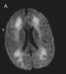
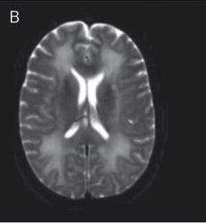
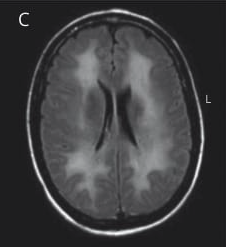
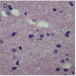
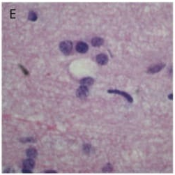
A 45-year-old female with a history of opioid use (morphine, oxycodone, heroin) presents with a 1-month history of altered mental status, bizarre behavior, and progressive cognitive decline. She had used opioids 2 months prior for 1 week, followed by a head injury 3 weeks later. Symptoms worsened over 1 month, including apraxia, aphasia, selective mutism, hyperreflexia, and wide-based gait. MRI brain showed bilateral white matter changes, and brain biopsy confirmed astrogliosis and microglial activation. The patient deteriorated rapidly and died 7 weeks after presentation.

Logo

medical imaging innovation in detection & diagnostic education

Get in touch
  • (832) 660-1111
  • info@imagecme.com

© 2026 - Image CME . All Rights Reserved

Share
  • WhatsApp
  • LinkedIn
  • Facebook
  • X
  • Email