Logo
Login Signup
images
X Ray
Chest X Ray showed a lobulated anterior mediastinal mass (yellow arrow) with mild elevation of the left hemidiaphragm (black arrow).
CT Scan
CT scan demonstrated a lobulated, left-sided anterior mediastinal mass (yellow arrow) and a 1-cm left-sided pleural nodule (white arrow).
View Discussion
  • Title: Myasthenia Gravis with Thymoma and Pleural Metastasis_
  • Disease Specialty: Oncology
  • Ease of Diagnosis: Difficult
  • Certainty: Certain
  • Ethnicity: Asian
  • Segment: Adult Female
  • Clinical Examination: Worsening dyspnea, poor PFTs, lobulated anterior mediastinal mass.
  • Patient Age: 67 Year
  • Patient Gender: Female

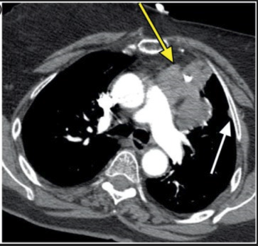
A 67-year-old woman with a 6-year history of myasthenia gravis presents with worsening exertional dyspnea. Past medical history includes coronary artery disease (stent placement), diabetes, and intermittent shortness of breath with previous oxygen dependence due to MG. Family history is notable for a sister who died from myasthenia gravis complications. She has a 60 pack-year smoking history, quit 10 years ago. Diagnostic findings include a chest radiograph showing a lobulated anterior mediastinal mass with mild elevation of the left hemidiaphragm (suggestive of phrenic nerve involvement) and a CT chest revealing an 8-cm left-sided anterior mediastinal mass with a 1-cm left pleural nodule consistent with drop metastasis.

Logo

medical imaging innovation in detection & diagnostic education

Get in touch
  • (832) 660-1111
  • info@imagecme.com

© 2026 - Image CME . All Rights Reserved

Share
  • WhatsApp
  • LinkedIn
  • Facebook
  • X
  • Email