Logo
Login Signup
images
CT Scan
A barium esophagram showed an irregular mass in the distal esophagus.
CT scans showed thickening of the distal esophagus.
an enlarged lymph node in the region of the gastrohepatic ligament, extending to the celiac axis.
PET Scan
The distal esophagus was intensely FDG avid Pet scanning (SUV 6.2).
The lymph node was also intensely FDG avid (SUV 8.3)
View Discussion
  • Title: Esophageal Cancer with Celiac Lymph Node Metastases
  • Diagnosis Title: Esophageal Adenocarcinoma with Lymph Node Involvement
  • Disease Specialty: Oncology
  • Ease of Diagnosis: Difficult
  • Certainty: Certain
  • Ethnicity: Asian
  • Segment: Adult Male
  • Image Quality: Medium
  • Clinical Examination: 45-year-old man with hematemesis, fungating distal esophageal mass, Barrett mucosa, T3N1Mx staging.
  • Patient Age: 45 years
  • Patient Gender: Male

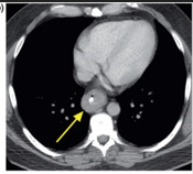
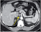
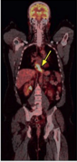
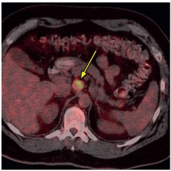
A 45-year-old man with a history of gastroesophageal reflux and hiatal hernia presented with 3-week history of hematemesis. Endoscopy revealed a fungating mass in the distal esophagus, biopsy shows poorly differentiated invasive adenocarcinoma arising in Barrett mucosa. Imaging findings include a barium esophagram showing an irregular distal esophageal mass and small hiatal hernia.

Logo

medical imaging innovation in detection & diagnostic education

Get in touch
  • (832) 660-1111
  • info@imagecme.com

© 2026 - Image CME . All Rights Reserved

Share
  • WhatsApp
  • LinkedIn
  • Facebook
  • X
  • Email