Logo
Login Signup
images
CT Scan
CT (brain window) red arrows show optic nerve sheath calcifications; green arrows show normal posterior fossa.
CT (bone window) – red arrows highlight optic sheath calcifications; green arrows show normal brain tissue.
Coronal CT – red arrows mark optic nerve sheath calcifications.
Abdominal CT – white arrows indicate CKD renal changes.
View Discussion
  • Title: Bilateral Optic Nerve Sheath Calcifications (CT scan).
  • Diagnosis Title: Secondary Hyperparathyroidism with Orbital Calcification in CKD.
  • Disease Specialty: Nephrology
  • Ease of Diagnosis: Difficult
  • Certainty: Certain
  • Ethnicity: Asian
  • Segment: Adult Male
  • Image Quality: Medium
  • Clinical Examination: Bilateral vision loss (amaurosis), normal fundoscopy, CT shows optic nerve sheath calcifications.
  • Patient Age: 66 Year
  • Patient Gender: Male

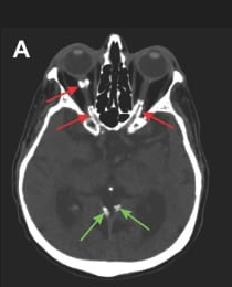
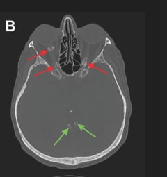
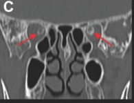
A 66 year old man with CKD on hemodialysis presented with bilateral amaurosis for 2 months. Fundoscopy was normal. CT revealed bilateral optic nerve sheath calcifications. Labs: PTH 105 pg/mL, Ca 12.5 mg/dL, PO₄ 3.8 mg/dL, ALP 192 U/L, 25-OH vitamin D 17 ng/mL. Suggests CKD-related mineral and bone disorder with rare orbital manifestation. Differentials: meningioma, glioma, phthisis bulbi, prior hemorrhage.
 

Logo

medical imaging innovation in detection & diagnostic education

Get in touch
  • (832) 660-1111
  • info@imagecme.com

© 2026 - Image CME . All Rights Reserved

Share
  • WhatsApp
  • LinkedIn
  • Facebook
  • X
  • Email